Understanding Esophagrams, Upper GI and Small Bowel Series for Your Child
Learn about esophagrams, upper GI and small bowel series and what to expect during these imaging procedures to help diagnose issues with your child’s digestive system.
X-rays are incredibly powerful tools that allow physicians to look inside the human body. By using little electromagnetic waves, they create images of your child’s bones, tissues and organs, which makes diagnosing and treating injuries and illnesses easier and more effective.
From examining a broken leg to diagnosing pneumonia in the lungs, X-rays can be used from head to toe for many purposes. If your child’s physician needs a closer look at their esophagus, stomach or small intestine, they may need one of the following digestive health tests:
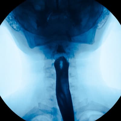
An esophagram, which takes pictures of their esophagus
An upper GI, which takes pictures of their esophagus, stomach and the connection between their stomach and small intestine
A small bowel series, which takes timed images of their small intestine
What Happens During an Esophagram, Upper GI or Small Bowel Series?
Though these tests take pictures of different parts of your child’s body, they’re performed in a similar way. During an esophagram, upper GI or small bowel series, the following steps will occur:
A Child Life specialist will meet with you and your child to help explain the procedure so your child can understand it, show you pictures of the room the procedure will occur in, and help your child develop a positive coping plan.
A radiology tech will be in the room (called the fluoro room) during the procedure. Two adults are allowed to accompany your child to the exam room. However, both caregivers will need to step behind the lead glass window during the procedure. If you’re pregnant, you will be unable to stay during the procedure. If you want to bring other children, make sure you bring one adult to stay in the waiting area with them.
Once you and your child are in the fluoro room, your child will need to change out of all of their clothes and into a hospital gown. You can help your child onto the exam table, where they will lie on their back.
Before the radiologist takes the X-ray, your child will need to drink a milkshake-like drink that contains Barium, which enables the doctor to see the structures of their digestive system on the images.
The radiologist will take X-rays of your child in different positions, such as on their back or on their side.
The entire procedure will take approximately 45 minutes for the esophagram and upper GI and up to 6 hours for the small bowel series.
How Can I Prepare and Support My Child for These Exams?
If your child needs an esophagram, upper GI or small bowel series, they may feel anxious about the procedure. This is completely normal, and there are ways to make them feel more comfortable, such as:
Using developmentally appropriate words to explain to your child what will happen, such as explaining the barium drink as either a Kool-Aid fruity drink or chocolate-flavored malt
Bringing a comforting item, such as a blanket or stuffed animal, for your child to have with them during their scan
Participating in One Voice, which is an initiative to promote a calming environment by limiting the number of voices in the room so your child knows who to focus on, such as a parent or procedural staff professional
Displaying a calm demeanor
Meet with one of our Child Life specialists — experts who can explain the procedure in child-friendly terms and help you and your child understand what will happen at every stage of the test
Questions?
Contact the Radiology Child Life specialist at 402.955.4042 or the Radiology Department at 402.955.5602.