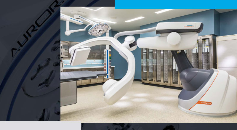
In less than four seconds, the massive robotic arm does a smooth 360-degree spin around the operating table, capturing precise 3D images of a baby’s heart that is the size of a walnut. This cutting-edge choreography has the power to unlock answers to some of the most complex pediatric cardiac cases, and it has made its debut in Omaha.
“State-of-the-art” seems like an understatement; Children’s Nebraska’s new heart catheterization labs boast a robotic imaging system that is the first of its kind in a pediatric setting worldwide. Manufactured by Siemens Healthineers, they call it the ARTIS pheno. Because Children’s is the first pediatric hospital to take advantage of the technology, its specialists will be defining the techniques for labs that follow.
“We’re positioning ourselves to become one of the premier programs in the country,” explains Jeff Delaney, M.D., Children’s director of interventional cardiology. “We’ve had a good reputation for the last 25 years, and now we’re also recognized not just as a place that does good work, but as a real leader with an exciting program.”
For the very smallest and sickest hearts (and the hearts of those who love them), the new labs are a global game-changer.
We’re positioning ourselves to become one of the premier programs in the country.
Jeff Delaney, M.D. Director of Interventional Cardiology
“Each of our patients has unique cardiac anatomy, and some of them are extraordinarily small. We’re doing extremely delicate maneuvers inside of the tiniest hearts,” says Dr. Delaney. “This system allows us to do this in a way that’s safer and better for children.”
Catheterizations are procedures used to visualize, diagnose and treat cardiovascular conditions, in which a long thin tube (catheter) is inserted into the body and then threaded through the blood vessels to the heart. Children’s team completes around 500 heart catheterizations each year; each procedure can last between two to six hours.
The new interventional catheterization suites also double as full-service operating rooms if and when surgery is needed. When images are needed, the robotic C-arm X-ray system, docked in one corner of the room, rises to glide up, over and around the procedure table.
What makes the new imaging equipment so special? According to Dr. Delaney, doctors used to have to constantly move the camera and adjust the angle to take the right pictures. Now, he says, “we can align an active fluoroscopic image to a previously obtained MRI image, CT scan or echocardiogram, and just spin it until it is right where we want it, hit a button and the camera moves to the exact place.”
Its hyper efficiency over a standard X-ray imaging system translates to better outcomes and a better experience for patient families.
“This technology gives us outstanding image quality at a faster speed, and if you can image at a faster speed, you’re using less radiation and less contrast and getting more information more quickly,” explains Dr. Delaney. “Many of our young patients have heart disease of the greatest severity and may need multiple catheterizations over the course of their lifetime, so this helps keep the cumulative dose of radiation much lower.”
You can hear the passion rise in Dr. Delaney’s voice when he describes the “roller coaster” that his heart patients and families often experience on their medical journeys. His eyes light up sharing how this innovation will help them.
“I want families in our community to know that we’re invested in their children and we’re taking it to the next level. This is absolutely what they can expect from us – nothing but the best.”


